
When it comes to the dental world, terminology and reasoning for dental work is new to patients. What’s the difference between a filling, onlay and a crown? Why do I need any of those? What causes a root canal and why do I need one? With some visual representation and x-rays we can breakdown the issue and provide a solution. Now let’s sink our teeth in!
 In our example here, the white appearance is a filling that is already placed on this tooth. Along the distal (the side away from the center of the mouth) surface, you can see a thin black shadow following underneath the filling. This is decay that is accumulated underneath the filling. If this decay continues and breaches more surfaces or into the nerve, this patient will be need a crown or potentially a root canal and crown. Luckily, the patient only needed their filling replaced to remove the decay.
 At our office, in addition to x-rays, we also take intra-oral pictures, which are pictures inside your mouth. What this picture is showing is decay that is accumulating along the grooves of the tooth. Decay like this is hard to typically see x-rays since they are two dimensional. Since they are 2D, they often don’t show decay on the chewing surface of teeth like your molars (back teeth). Pictures like this are helpful to show insurance companies why certain dental work was needed and why.
 Here we see decay that has reached the DFL surfaces which are Distal (toward the back of the mouth), Facial (toward the front of the face), and Lingual (toward the tongue). As the area of decay is small, a resin filling is the only dental work needed at the time to remove the decay and protect from future issues.
 Tooth #14 on this x-ray has a very large restoration on it, commonly known as a filling. Beneath this filling, we can see shading (which indicates decay) all along the filling. The decay here has reached the MOL surfaces, Mesial (toward center of mouth), Occlusal (chewing surface of mouth) and the Lingual (toward the tongue). After removal of the filling and clearing out of the previous restoration, it became clear that a filling was no longer appropriate for this situation. Why? Now let’s look at our intra-oral picture that we took below for the answer.
 This area is too large for a filling, it would not be able to stand the intense force we use with our back teeth without fracturing or simply coming out. This situation is perfect for is an onlay. An onlay is made out of pure porcelain, is more extensive than a filling, but less than a crown. This type of restoration is more dependable when it comes to covering more cusps as it is a stronger material made for these situations. Once bonded in, the onlay is ready to go and function.
 When old fillings take over 50% of the tooth and begin to chip, fracture, fall out and/or gather decay, it is appropriate to crown the tooth. Crowns at Goldstein Dental are made in-office out of pure porcelain by our CEREC CAD/CAM machine. Here we see a very large restoration that also has decay beneath it. On the Mesial surface (toward the middle of the mouth) we can see that there is an actual space between the filling and natural tooth. That is called an ‘open margin’ which allows decay to enter with direct access to the natural tooth and can even become a food trap. The patient was experiencing hot/cold sensitivity and general sensitivity while chewing. This tooth was crowned (within an hour) and our patient was on their way to enjoy their day.

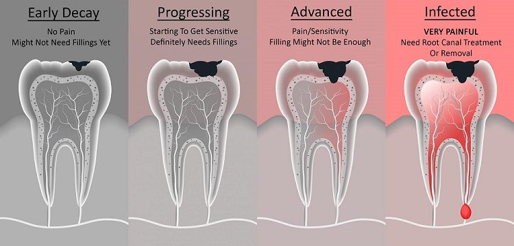
This illustration shows an idea of how it is decided on what treatment is generally needed. With early decay, the decay has breached the enamel. A patient may start showing signs of sensitivity and a filling can be placed to rectify this. Once the decay progresses and breaches the dentin, sensitivity ensues and a filling is needed. As the decay continues, it starts to attack more surfaces and reach deeper into the tooth. At this stage, an onlay may be necessary or if the decay has wrapped around a tooth, a crown will be necessary. In the infected picture, the decay has reached the pulpitis and is attacking the nerve of the tooth. This is no longer sensitive, it is painful. Swelling and throbbing can begin to occur, infection is present and can appear in the gums by the end of the root of the tooth, and the pain can be such that it will wake patients up at night. At this point in time, a root canal is necessary. The root of the tooth (and infection) are removed and cleared out. A permanent filling (referred to as a Post & Core) is placed in place of the root to prevent future infection. Finally, a crown is placed on the top of the tooth to completely seal the tooth.